Cell Division Zebrafish Embryo
Time-lapse confocal microscopy of asymmetric cell division in the brain of a live zebrafish embryo. Neurogenesis in the developing central nervous system consists of the induction and proliferation of neural progenitor cells and their subsequent differentiation into mature neurons.
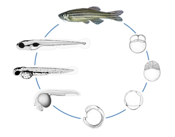
 Zebrafish Development Embryology
Together these results suggest that the environment of the zebrafish embryo supports survival of human metastatic melanoma cells over several days and that these cells in the zebrafish are able to change cellular morphology migrate and undergo cell division.

Cell division zebrafish embryo. Using quantitatively measured 4D live-imaging data features of V2 cell-shape at each time point prior to division were extracted and a statistical model. Animal pole view of 2-3 hours post fertilization zebrafish embryos injected with PCNA-GFP protein. For example using a confocal imaging technique we have shown that a localized Ca2 elevation was clearly associated with the onset of cytokinesis in zebrafish embryo Chang and Meng 1995 J.
One consequence of aberrant mitoses in cfy embryos is an increase in cell death. The zebrafish curly fry cfy mutation leads to embryonic lethality and abnormal cell divisions starting at 12-14 h postfertilization hpf during neural tube formation. The results show the incorporation of BrdU into the newly synthesized DNA in the human melanoma cell nuclei Fig.
Studies of the developing zebrafish embryo have revealed similarities to the early cell divisions of other vertebrates such as Xenopus. In contrast to the fate of melanoma cells human melanocytes transplanted into zebrafish embryos most frequently become distributed to their normal microenvironment of the. Chemicals found in our zebrafish system expand postnatal muscle satellite cells from mice and specify myogenic differentiation from human pluripotent cells thus helping to address two of the more vexing challenges in the production of mammalian muscle precursors for experimental applications and ultimately cell.
Using intrinsic second- and third- harmonic generation SHG THG signals from mitotic spindles and cell. We choose V2 neural progenitor cells in developing zebrafish embryo as their successive shape changes can be visualized in real-time in vivo. This finding was later confirmed in studies using aequorin as a Ca2 probe.
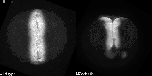
The mid blastula transition MBT ensues during the tenth cell division which is approximately 3 hpf. A noteworthy attribute of the zebrafish embryo culture system is its ability to identify pathways that influence tissue specification and progenitor cell expansion across species. The data show that human metastatic melanoma cells placed in the zebrafish embryo survive exhibit motility and divide.
This image sequence shows a single neural progenitor a cell which can divide and differentiate into a limited number of neural cell types dividing to produce two daughter cells. In homozygous mutant embryos mitoses are disorganized with an increase in mitotic figures throughout the developing neural tube. We developed a relatively simple mathematical method of describing cellular geometry of V2 cells to predict cell division-timing based on their successively changing shapes in vivo.
In the last 20 years the zebrafish has proven to be an excellent model organism to study neurogenesis in the embryo. External as well as internal cues orchestrate neurogenesis in a precise temporal and spatial way. Following the dynamic process of cell proliferation in whole embryos.
More likely however DHX8 is required to splice pre-mRNA molecules that encode proteins directly required for mitotic exit. Our results in HeLa cells confirmed the observation that loss of dhx8 in the zebrafish led to severe cell division defects. The sequence starts at the 8 oclock position on a clock face with the two daughter cells sitting side by side on.
PCNA-GFP appears in the nucleus during S-phase. Cells treated with DHX8 shRNA were unable to complete mitosis correctly leading to multi-nucleated cells and cell death. 3E arrow thus corroborating cell division within the zebrafish embryos.
We therefore turned to zebrafish embryos whose external development and transparency provide good access to almosteverystageofembryogenesisUsingthehspa8promoter we generated transgenic zebrafish lines that express mAG-hGem1110 or mKO2-hCdt130120. The mitotic defect is seen in a variety of tissues including the central nervous system CNS. The melanoma cells do not form tumors nor integrate into host organs but instead become scattered throughout the embryo in interstitial spaces reflecting the dedifferentiated state of the cancer cells.
It is possible that DHX8 is directly required for mitotic exit thus suggesting a new function for the gene. Recent studies suggested that a Ca2 signal is involved in the regulation of cell division. That provides in toto quantitative imaging of cell divisions in unstained zebrafish embryos during the first 10 division cycles.
MBT is accompanied by loss of. In the zebrafish the first seven cell divisions are synchronous and cycle rapidly between DNA replication S phase and mitosis M phase without the intervening gap phases G1 and G2 Kimmel et al 1995. Here we used a 4-D confocal measurement technique to further characterize the properties.
Optimized scanning along circular trajectories is used to preferentially deliver the excitation energy to the innermost regions of the spherical embryo. Time-lapse imaging of PCNA-GFP allows for live.
 Zebrafish Cell Culture
 Axis Specification In Zebrafish Is Robust To Cell Mixing And Reveals A Regulation Of Pattern Formation By Morphogenesis Sciencedirect
 Scientifically Speaking Zebrafish Development From A Few Cells To An Embryo
 Illuminating Cell Cycle Progression In The Developing Zebrafish Embryo Pnas
 Visualizing Enveloping Layer Glycans During Zebrafish Early Embryogenesis Pnas
 Spatio Temporal Mrna Dynamics In The Early Zebrafish Embryo Biorxiv
Https Www Sciencedirect Com Science Article Pii S0091679x08618178 Pdf Md5 81b49f67f8289863678aee9d836f2e79 Pid 1 S2 0 S0091679x08618178 Main Pdf
 Schematic Depiction Of Epiboly Initiation And Progression In The Download Scientific Diagram
 How To Plumb A Pisces Understanding Vascular Development And Disease Using Zebrafish Embryos Developmental Cell
 Shaping The Zebrafish Myotome By Intertissue Friction And Active Stress Pnas

High Throughput Screening For Bioactive Molecules Using Primary Cell Culture Of Transgenic Zebrafish Embryos Sciencedirect
 Dynamically Evolving Cell Sizes During Early Development Enable Normal Gastrulation Movements In Zebrafish Embryos Biorxiv
 Zebrafish Early Embryo Sorting Bionomous
 Revealing The Mysteries Of Early Development Washington University School Of Medicine In St Louis

 Cell Proliferation Patterns In Early Zebrafish Development Mendieta Serrano 2013 The Anatomical Record Wiley Online Library
 Single Cell Reconstruction Of Developmental Trajectories During Zebrafish Embryogenesis Science
 Plk1 And Plk4 Mediated Asymmetric Mitotic Centrosome Size And Positioning In The Early Zebrafish Embryo Biorxiv